Procedure explained · Nose and airways
Rhinoscopy explained: looking inside the nose
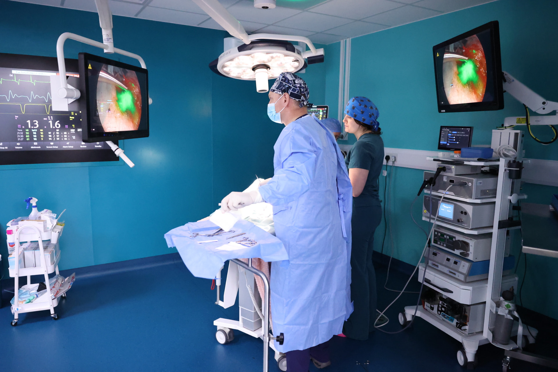
Rhinoscopy is an examination of the inside of the nose using a small camera, carried out under general anaesthetic. It lets the vet look directly at the nasal passages, search for foreign material or abnormal tissue, and take samples. It is usually one part of an investigation, often alongside a CT scan.
Draft — awaiting clinical review. This guide is an introductory draft. It hasn’t yet been approved by a named clinical reviewer and is hidden from search engines until it has.
What is rhinoscopy?
The word comes from rhino- (nose) and -scopy (looking). A thin camera, called an endoscope, is passed into the nose so the vet can see the lining on a screen in magnified detail.
There are usually two parts. The front of the nose is examined through the nostrils, often with a slim, rigid camera. The space at the back of the nose — above the soft palate — is examined with a flexible camera that bends around from the mouth. This back area is a common hiding place for foreign objects and growths.
Image placeholder
Rhinoscopy camera routes
Why might a vet recommend it?
Rhinoscopy may be suggested when signs such as persistent sneezing, nasal discharge, nosebleeds or noisy breathing haven't been explained by an examination and simpler tests. It can help to:
- Look for, and sometimes remove, foreign objects such as grass seeds
- See areas of inflammation, fungal plaques or abnormal tissue
- Take small tissue samples (biopsies) for a pathologist to examine under a microscope
- Collect swabs or flush samples for laboratory testing
How rhinoscopy and CT work together
CT and rhinoscopy answer different questions. CT shows the shape and structure of the nose, sinuses and surrounding bone, including areas a camera can't reach. Rhinoscopy shows the surface of the lining directly and allows targeted samples.
When both are planned, CT is usually done first, often during the same anaesthetic. Taking samples can cause bleeding, which could make the scan harder to interpret — and the scan can help the vet decide where to look and sample.
What to expect on the day
Arrangements differ between practices and referral centres, so always follow the instructions you're given. In general:
- You'll usually be asked not to feed your pet for a set time beforehand
- Your pet will be examined, and blood tests may be done to check they're well enough for anaesthesia
- The procedure is carried out under general anaesthetic, with your pet monitored throughout
- Your vet will usually call you once your pet is recovering
- Many pets go home the same day, but this varies with the individual and what was done
Video placeholder
Explained video: a rhinoscopy, step by step
Afterwards
After samples are taken, it's common for a pet to sneeze more and to have a little blood-tinged discharge for a short time. Your vet will explain what's expected and what should prompt a call.
Laboratory results often take several days. Sometimes the findings give a clear answer; sometimes they narrow down the possibilities, and further discussion or treatment trials are needed.
A balanced view
Weighing it up: rhinoscopy
Every option has trade-offs. What’s right depends on your pet, and is best decided with your vet.
Potential benefits
- A direct, magnified view of the nasal lining without a surgical incision
- Samples can be taken from the areas that look most abnormal
- Some foreign objects can be removed during the same procedure
Risks
- A general anaesthetic is required, which always carries some risk
- Bleeding from the nose is common after samples and is usually mild, but can occasionally be more significant
- Temporary increase in sneezing or discomfort
Limitations
- Some areas, such as parts of the sinuses, can be difficult or impossible to see
- Blood or mucus can obscure the view
- Biopsies are small, and occasionally don't give a definitive answer
- It doesn't show what's happening within or beyond the bone — CT is better for that
Alternatives
- CT on its own, depending on the question being asked
- X-rays, which are more widely available but show less detail
- Treatment trials or monitoring, where appropriate
- Surgical exploration of the nose, in specific circumstances
Questions to ask your vet
You might find it helpful to note these down before your appointment.
- 01What are you hoping rhinoscopy will show for my pet?
- 02Will a CT scan be done as well, and in what order?
- 03Will samples be taken, and when should I expect results?
- 04What is normal after the procedure, and what should prompt me to call?
- 05If nothing is found, what would the next step be?
References
References will be added here following clinical review. We only list sources that have been checked.
Who wrote and reviewed this
- Written by
- To be added once verified
- Clinically reviewed by
- To be added once verified
- Review date
- To be added once verified
Pet Care Explained is produced by Vetemis Referrals. Learn about investigations at Vetemis(opens vetemis.co.uk)
Related guides
My dog keeps sneezing: what might my vet investigate?
Why dogs sneeze, when to contact your vet, and which tests might be considered if sneezing doesn't settle.
CT, ultrasound and endoscopy: why might different tests be needed?
A plain-language comparison of three common investigations, and why your vet might suggest more than one.
Endoscopy explained: what can a camera examination tell us?
How a camera examination of the gut works, what it can reveal, and the questions it can't answer on its own.