Tests compared · Nose and airways
CT, ultrasound and endoscopy: why might different tests be needed?
Each test answers different questions. Ultrasound looks at soft organs in real time; CT builds a detailed three-dimensional picture, including bone and air-filled spaces; endoscopy looks directly at the inside surface of hollow organs and allows samples to be taken. Vets often combine them because no single test shows everything.
Draft — awaiting clinical review. This guide is an introductory draft. It hasn’t yet been approved by a named clinical reviewer and is hidden from search engines until it has.
A simple way to think about it
Imagine trying to understand a problem in a town. A CT scan is like a detailed 3D map: it shows where everything is and how it fits together. Ultrasound is like a live view from a drone: you can watch things move and look closely at particular buildings. Endoscopy is like walking down the street: you see the surfaces right in front of you in fine detail, and can pick things up.
Each view is useful. None tells the whole story on its own.
Ultrasound
Ultrasound uses sound waves to create a moving picture of soft tissues. A probe is placed on the skin, usually after clipping a small patch of fur.
- Good for: organs in the abdomen such as the liver, kidneys, bladder and intestines; measuring the thickness of the gut wall; looking at the heart; guiding needle samples
- Limited by: air and bone, which block sound waves — so it's less useful for the lungs, nose and skull
- Usually: done with your pet awake or lightly sedated
CT (computed tomography)
CT uses X-rays taken from many angles, combined by a computer into detailed cross-sectional and 3D images. A contrast agent may be injected to highlight blood vessels and certain tissues.
- Good for: the nose and sinuses, skull, chest and lungs, bones and joints; seeing the extent of a problem; planning surgery
- Limited by: it shows structure, not the surface of a lining; it can't always tell what a change is without samples
- Usually: needs your pet to be completely still, so is done under general anaesthetic or heavy sedation
Endoscopy
Endoscopy uses a camera to look inside hollow organs such as the gut, airways, nose or bladder.
- Good for: seeing the lining directly; taking targeted samples; removing some foreign objects
- Limited by: it can't see through walls or into solid organs, and some areas are out of reach
- Usually: performed under general anaesthetic
At a glance
| Test | Particularly useful for | Main limitations | Sedation or anaesthetic |
|---|---|---|---|
| X-rays | Bones, chest, some foreign objects and stones | Overlapping structures; less soft-tissue detail | Sometimes sedation |
| Ultrasound | Abdominal organs, gut wall, guiding samples | Blocked by air and bone | Often awake or light sedation |
| CT | Nose, skull, chest, bones; 3D planning | Shows structure, not surface lining | Usually anaesthetic or heavy sedation |
| Endoscopy | Inner lining; targeted samples; some object removal | Can't see beyond the lining | General anaesthetic |
Why more than one test may be needed
Tests are often used in a sequence, with each result shaping the next step. For example, a CT scan of the nose might show where an abnormal area is; rhinoscopy can then be used to look at it and take samples. In the abdomen, ultrasound may show a thickened area of gut, and endoscopy or surgery may be used to sample it.
Sometimes tests are combined in one anaesthetic to reduce the number of anaesthetics your pet needs.
Making decisions together
It's reasonable to ask how each test would change the plan for your pet. Factors that are often worth discussing include your pet's general health, the need for anaesthesia, cost and insurance, travel and how much information is likely to be gained.
A balanced view
Weighing it up: choosing between investigations
Every option has trade-offs. What’s right depends on your pet, and is best decided with your vet.
Potential benefits
- Choosing the right test for the question can avoid unnecessary procedures
- Combining tests can give a more complete picture
- Planning tests together can reduce the number of anaesthetics
Risks
- Sedation and anaesthesia carry some risk
- Contrast agents used in CT can occasionally cause reactions
Limitations
- No single test shows everything
- Imaging often can't confirm exactly what a change is without samples
- Some results are inconclusive and need further discussion
Alternatives
- Starting with simpler tests such as blood tests or X-rays
- Monitoring or treatment trials where appropriate
- Surgical exploration in specific situations
Questions to ask your vet
You might find it helpful to note these down before your appointment.
- 01What question are we trying to answer with this test?
- 02How would the result change what we do next?
- 03Could any tests be done during the same anaesthetic?
- 04Are there simpler tests we should try first?
- 05What would you do if the result is unclear?
References
References will be added here following clinical review. We only list sources that have been checked.
Who wrote and reviewed this
- Written by
- To be added once verified
- Clinically reviewed by
- To be added once verified
- Review date
- To be added once verified
Pet Care Explained is produced by Vetemis Referrals. Learn about investigations at Vetemis(opens vetemis.co.uk)
Related guides
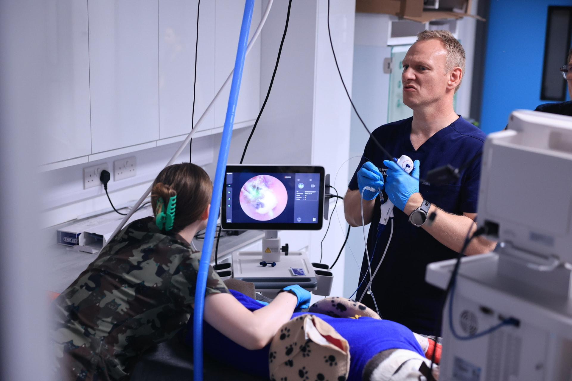
Endoscopy explained: what can a camera examination tell us?
How a camera examination of the gut works, what it can reveal, and the questions it can't answer on its own.
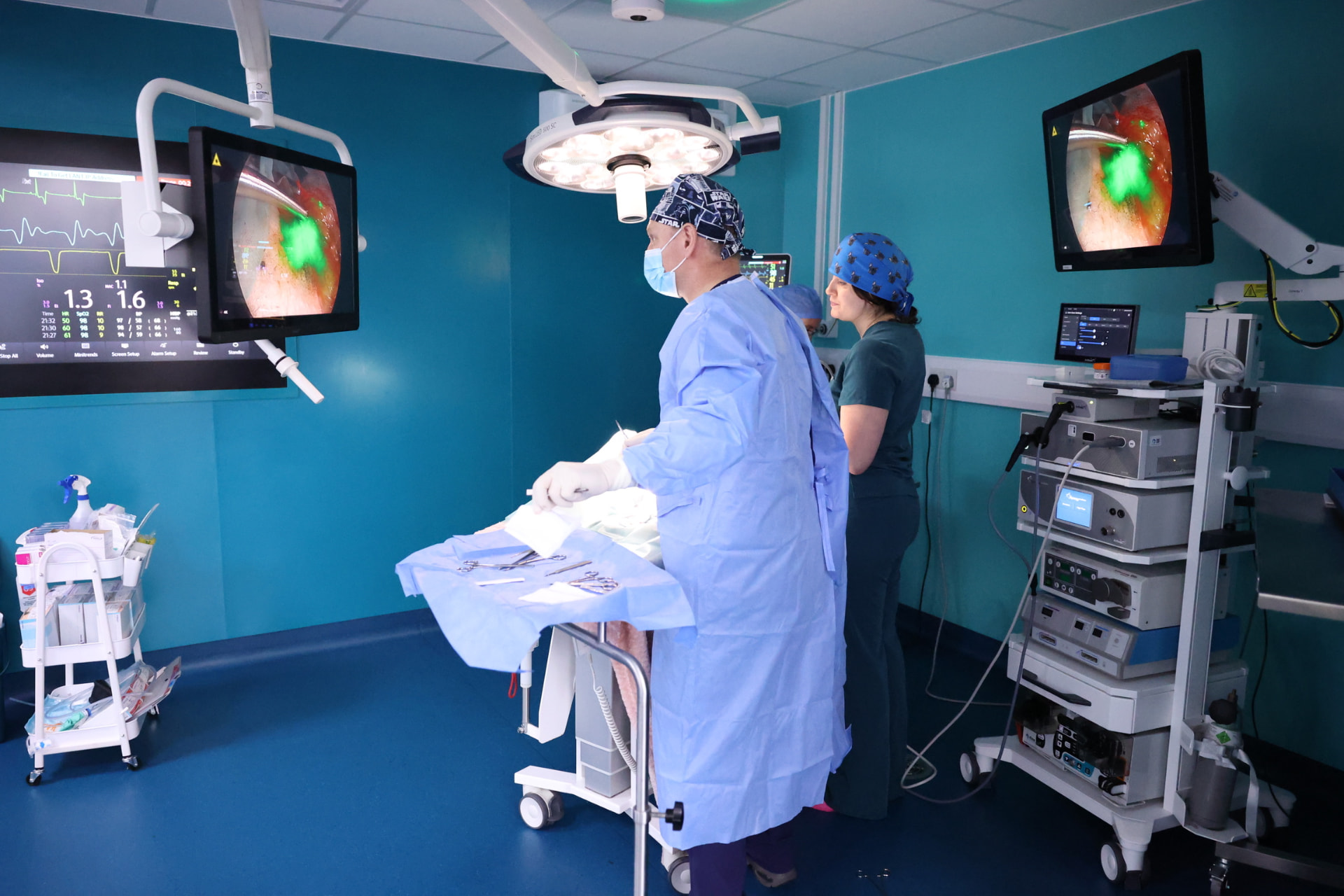
Rhinoscopy explained: looking inside the nose
What happens during a camera examination of the nose, what it can and can't show, and what to expect on the day.
My dog keeps sneezing: what might my vet investigate?
Why dogs sneeze, when to contact your vet, and which tests might be considered if sneezing doesn't settle.