Procedure explained · Digestion
Endoscopy explained: what can a camera examination tell us?
Endoscopy uses a slim, flexible tube with a camera and light to look inside hollow organs — most often the gullet, stomach and first part of the small intestine. It can show the inner lining directly, allow small tissue samples to be taken, and sometimes remove swallowed objects without open surgery. It can't see through the wall of the gut or assess organs around it.
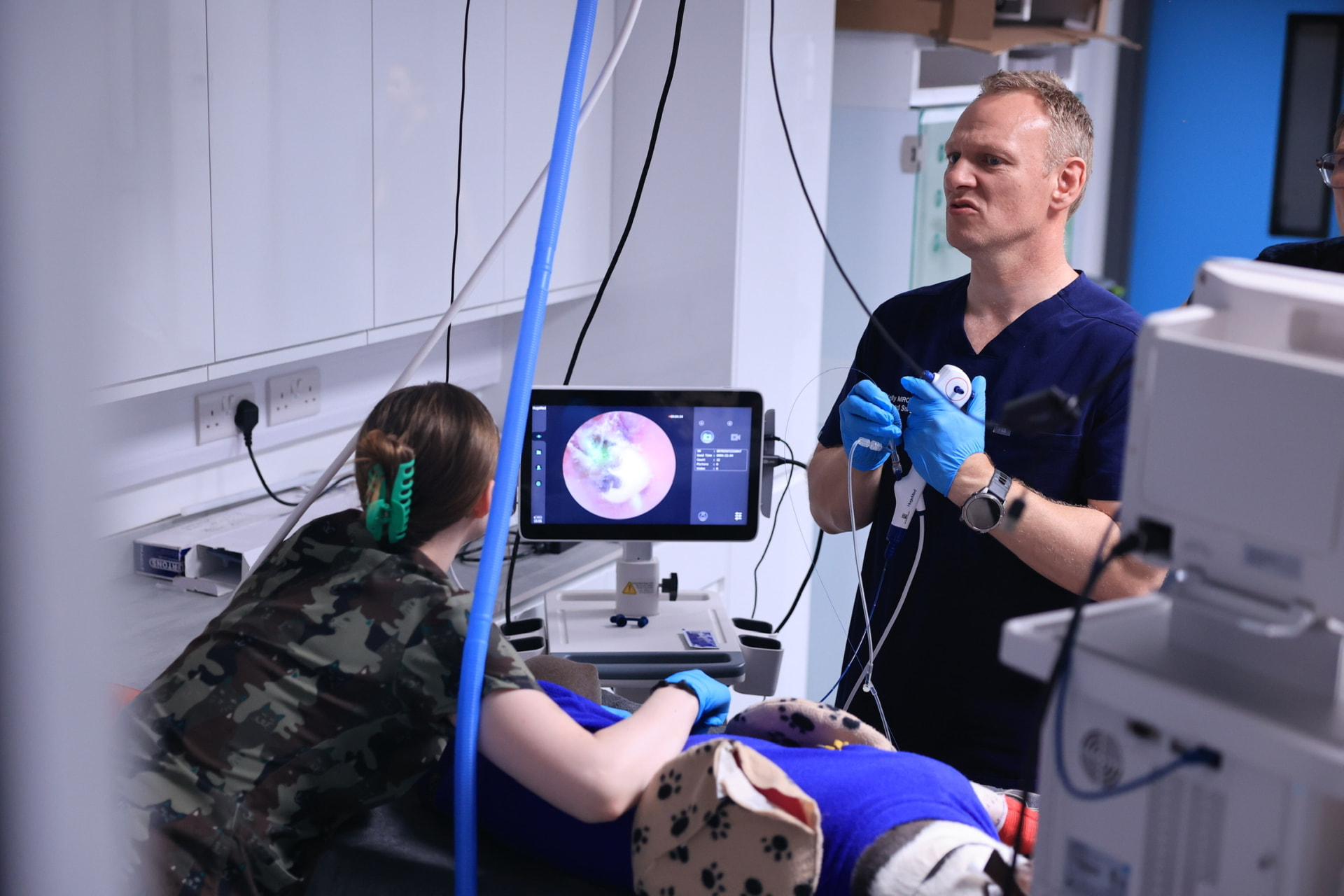
Draft — awaiting clinical review. This guide is an introductory draft. It hasn’t yet been approved by a named clinical reviewer and is hidden from search engines until it has.
What does 'endoscopy' mean?
Endo- means 'within' and -scopy means 'looking'. An endoscope is a camera on a long, flexible tube. It is passed through a natural opening — usually the mouth — so the vet can see the inside of the body on a screen.
The same idea is used in different parts of the body, and each examination has its own name:
- Upper gastrointestinal endoscopy
- The gullet (oesophagus), stomach and the first part of the small intestine (duodenum).
- Colonoscopy
- The large intestine, examined from the other end.
- Bronchoscopy
- The windpipe and airways of the lungs.
- Rhinoscopy
- The nasal passages.
- Cystoscopy
- The urethra and bladder.
Image placeholder
Digestive tract anatomy
What can endoscopy show?
Because the camera looks directly at the lining, endoscopy can reveal:
- Inflammation, redness or ulcers
- Swallowed objects, such as toys, bones or fabric
- Narrowing (a stricture) of the gullet
- Lumps or abnormal tissue on the inner surface
Very often, the most important part is taking small tissue samples (biopsies). A pathologist examines these under a microscope, which can show changes that look completely normal to the eye.
Removing swallowed objects
If an object is stuck in the gullet or stomach, it can sometimes be grasped and removed using instruments passed through the endoscope. This may avoid open surgery. Whether it's possible depends on the size, shape and position of the object, and how long it has been there.
What endoscopy can't tell us
- It only sees the inner surface — not the deeper layers of the gut wall
- It can't assess organs outside the gut, such as the liver, pancreas or lymph nodes
- Much of the small intestine is out of reach
- Biopsies are small and come from the inner layers, so some conditions can be missed
- A normal appearance doesn't rule out disease — laboratory results matter
Because of this, endoscopy is often combined with other tests. If deeper samples are needed, your vet may discuss surgical biopsies instead.
Why other tests often come first
Digestive signs have many possible causes, and lots of them can be investigated or managed without an anaesthetic. Depending on your pet, your vet may suggest blood tests, faecal tests, ultrasound, X-rays or a carefully managed diet trial before considering endoscopy.
Ultrasound is particularly useful alongside endoscopy, because it can show the thickness of the gut wall and the organs around it.
What usually happens on the day
- Your pet will need to have an empty stomach — follow the fasting instructions you're given
- Endoscopy in animals is performed under general anaesthetic
- The procedure itself is often relatively short, but timings vary
- Most pets recover quickly, although some may have a mildly sore throat
- Biopsy results usually take several days
Video placeholder
Explained video: a guided look inside the stomach
A balanced view
Weighing it up: gastrointestinal endoscopy
Every option has trade-offs. What’s right depends on your pet, and is best decided with your vet.
Potential benefits
- A direct view of the lining without an incision
- Biopsies can be taken without open surgery
- Some swallowed objects can be removed, which may avoid surgery
- Recovery is usually quick
Risks
- A general anaesthetic is always needed
- Rare but serious complications include bleeding or a tear (perforation) in the gut wall
- Mild throat irritation afterwards
Limitations
- Only the inner surface of reachable areas can be seen
- Biopsies may not capture deeper changes
- It doesn't assess organs outside the gut
Alternatives
- Ultrasound, X-rays or CT, depending on the question
- Blood and faecal tests
- Diet trials or medical treatment with review
- Surgical biopsies, by open or keyhole surgery, when deeper samples are needed
Questions to ask your vet
You might find it helpful to note these down before your appointment.
- 01What are you hoping endoscopy will show?
- 02Are there any tests we should do first?
- 03Will biopsies be taken, and when will results be available?
- 04If an object can't be removed with the endoscope, what happens next?
- 05What signs should I watch for after the procedure?
References
References will be added here following clinical review. We only list sources that have been checked.
Who wrote and reviewed this
- Written by
- To be added once verified
- Clinically reviewed by
- To be added once verified
- Review date
- To be added once verified
Pet Care Explained is produced by Vetemis Referrals. Learn about investigations at Vetemis(opens vetemis.co.uk)
Related guides
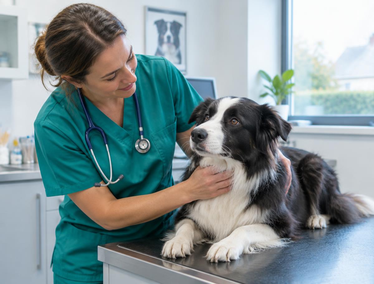
CT, ultrasound and endoscopy: why might different tests be needed?
A plain-language comparison of three common investigations, and why your vet might suggest more than one.
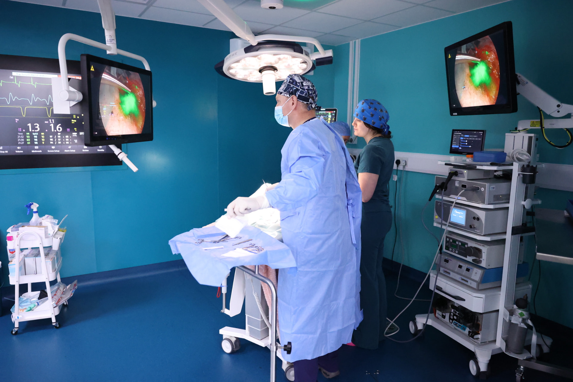
Rhinoscopy explained: looking inside the nose
What happens during a camera examination of the nose, what it can and can't show, and what to expect on the day.
Bladder stones: understanding investigation and treatment options
Signs of bladder stones, how vets investigate them, and the range of treatment options — from diet to surgery.