Condition guide · Bladder and urinary tract
Bladder stones: understanding investigation and treatment options
Bladder stones are hard collections of minerals that form in the urine. They can cause straining, frequent urination and blood in the urine — and sometimes a dangerous blockage. Treatment depends on the type, size and position of the stones and on the individual pet. Options range from special diets that can dissolve some stones, to removing them with camera-guided techniques or traditional surgery.
Draft — awaiting clinical review. This guide is an introductory draft. It hasn’t yet been approved by a named clinical reviewer and is hidden from search engines until it has.
When to contact your vet
Draft guidance awaiting clinical review. If you are unsure, always contact your vet — they can advise how urgently your pet should be seen.
Contact a vet immediately if
- Your pet is straining to urinate but producing little or no urine — this can mean a blockage and is an emergency, particularly in male cats and male dogs
- Crying, restlessness or obvious pain when trying to urinate
- Vomiting, weakness or lethargy alongside urinary signs
If your practice is closed, call their out-of-hours service.
Book an appointment with your vet if
- Blood in the urine
- Urinating more often, or in small amounts
- Accidents indoors, or licking around the genitals more than usual
What are bladder stones?
Vets may call them uroliths or calculi. They form when minerals in the urine come together into crystals, which can then build into stones ranging from sand-like grains to large single stones.
There are several types, named after what they're made of. Two of the most commonly seen in dogs and cats are struvite and calcium oxalate. Others include urate and cystine stones. The type matters because it affects which treatments are likely to work and how recurrence can be reduced.
Image placeholder
Urinary tract anatomy
Signs owners may notice
Some pets with stones show no signs at all, and the stones are found during tests for something else. Others may:
- Strain or take a long time to urinate
- Urinate more often, in small amounts
- Pass blood-tinged urine
- Have accidents in the house or urinate in unusual places
- Lick around the genitals
These signs aren't specific to stones. Urinary infections, inflammation of the bladder (especially in cats) and other conditions can look very similar, which is why testing matters.
How vets usually investigate
- Urine tests
- Look for blood, crystals, infection and concentration. A sample collected directly from the bladder with a fine needle may be suggested for culture.
- X-rays
- Many stones show up on X-rays, but some types are hard to see.
- Ultrasound
- Can show stones that X-rays may miss, and look at the bladder wall and kidneys.
- Blood tests
- Check kidney function and general health, and can sometimes give clues about why stones are forming.
- Stone analysis
- Once a stone is passed or removed, a laboratory can confirm exactly what it's made of. This guides prevention.
Treatment options
There isn't one best treatment for every pet. Your vet will consider the likely stone type, the number and size of stones, where they are, whether there's a blockage, your pet's sex and size, and what facilities are available.
- Dissolution diet
- Some stones — particularly struvite — can often be dissolved with a prescription diet, sometimes with medication. This needs regular check-ups and isn't suitable for all stone types or if there's a blockage.
- Flushing small stones (urohydropropulsion)
- Under anaesthetic, the bladder is filled with fluid and gently expressed so that small stones can pass out through the urethra.
- Cystoscopic removal
- A thin camera passed along the urethra lets the vet see the stones and remove smaller ones with a tiny basket. This is generally more practical in females, whose urethra is wider.
- Laser fragmentation (lithotripsy)
- A laser passed through the camera breaks stones into smaller pieces that can be removed. This requires specialist equipment and isn't widely available.
- Minimally invasive surgical removal (PCCL)
- Percutaneous cystolithotomy uses a small incision into the bladder and a camera to find and remove stones.
- Open surgery (cystotomy)
- The bladder is opened through an incision in the abdomen and the stones removed. This remains an effective and appropriate choice for many pets.
Image placeholder
Annotated image: how a cystoscope reaches the bladder
Reducing the chance of stones coming back
Some types of stone have a tendency to recur. Depending on the stone type, your vet may recommend a particular diet, ways to encourage your pet to drink more, regular urine checks and repeat imaging. Knowing the stone type from laboratory analysis helps make this plan as useful as possible.
A balanced view
Weighing it up: treatment options for bladder stones
Every option has trade-offs. What’s right depends on your pet, and is best decided with your vet.
Potential benefits
- Dissolution diets can avoid an anaesthetic for suitable stone types
- Camera-guided techniques can mean smaller or no incisions
- Open surgery is widely available and allows removal of many or large stones
Risks
- Any procedure under anaesthetic carries some risk
- Stones can move and cause a blockage, including during dissolution
- Surgical techniques carry risks such as bleeding, infection or urine leakage
Limitations
- Diets don't dissolve all stone types
- Cystoscopic removal is limited by stone size and the size of the urethra
- Specialist techniques aren't available everywhere
- Small fragments can sometimes be left behind, so follow-up imaging is often recommended
Alternatives
- Monitoring small stones that aren't causing problems, if your vet feels this is appropriate
- Combining approaches — for example diet followed by flushing
- Referral for specialist techniques, or open surgery in your own practice
Questions to ask your vet
You might find it helpful to note these down before your appointment.
- 01What type of stone do you suspect, and could it dissolve with diet?
- 02Is there any sign of a blockage, and what should I watch for at home?
- 03Which removal options are suitable for my pet, and why?
- 04Will the stones be sent for analysis?
- 05What follow-up will we need to reduce the chance of stones returning?
References
References will be added here following clinical review. We only list sources that have been checked.
Who wrote and reviewed this
- Written by
- To be added once verified
- Clinically reviewed by
- To be added once verified
- Review date
- To be added once verified
Pet Care Explained is produced by Vetemis Referrals. Learn about investigations at Vetemis(opens vetemis.co.uk)
Related guides
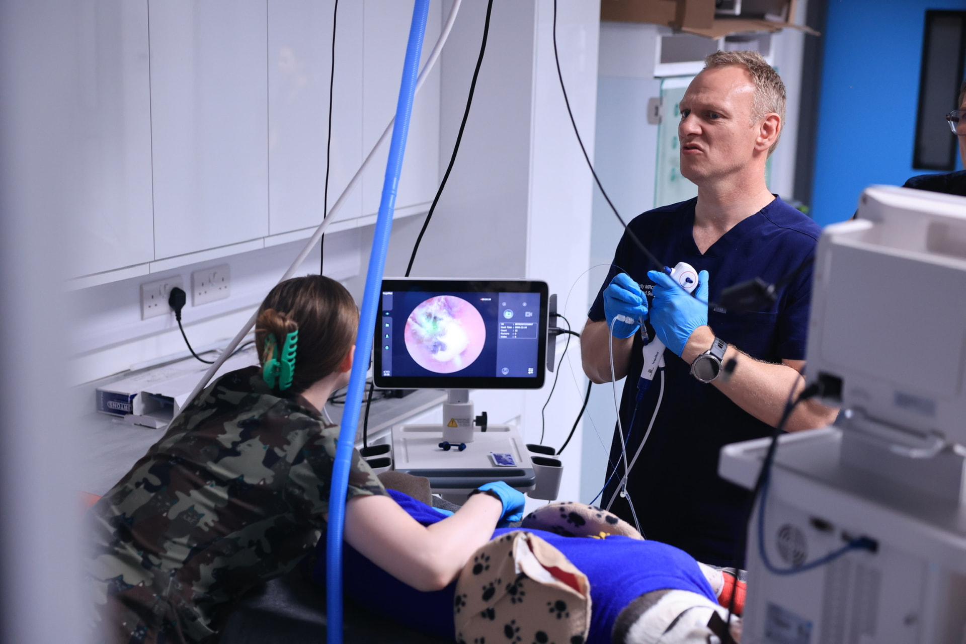
Endoscopy explained: what can a camera examination tell us?
How a camera examination of the gut works, what it can reveal, and the questions it can't answer on its own.
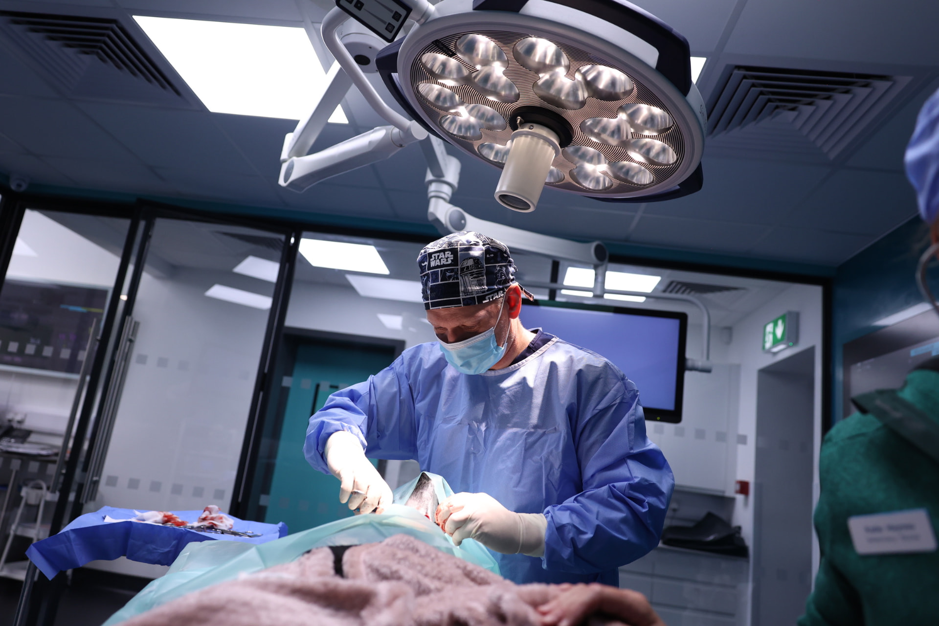
Keyhole spay explained: questions to ask your vet
How a keyhole spay differs from a traditional spay, its potential benefits and limitations, and what to ask.
CT, ultrasound and endoscopy: why might different tests be needed?
A plain-language comparison of three common investigations, and why your vet might suggest more than one.